
Automates and Organises Quality Assurance in Radiation Therapy and Medical Imaging

MOD PET
QUALIMAGIQ software module for a comprehensive and automatic quality control of a PET camera
Connected to the QUALIMAGIQ platform, it takes the MOD-PET module only 2 mouse clicks to analyse all of the DICOM images originating from comprehensive quality control of a positon emission gamma camera, which may or may not be operating in conjunction with an X-ray CT scanner (PET or PET-CT)
BY FAR THE BEST SOLUTION FOR PET CAMERA QA!
The MOD-PET module automates the tests proposed by the NEMA and SFPM given below:
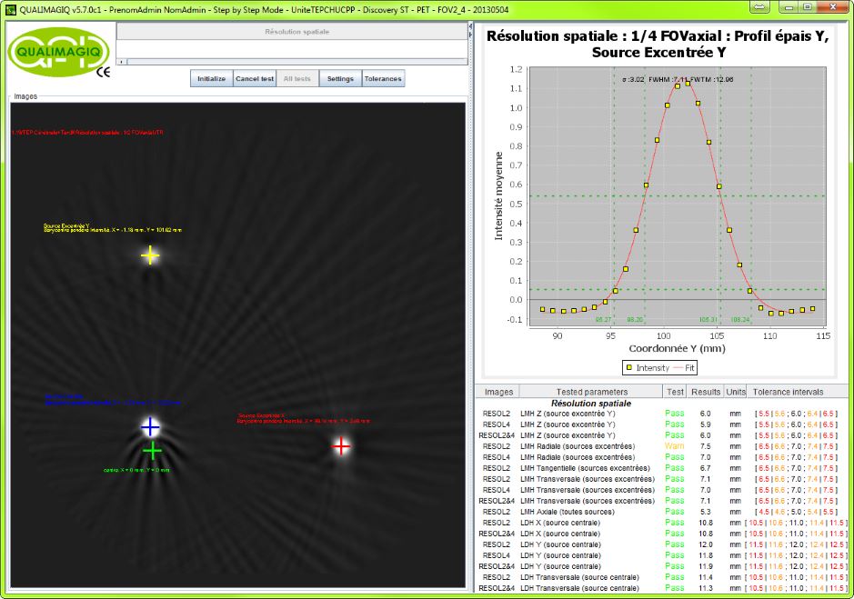
- Spatial resolutions1: FWHM and FWTM (Full Width at Half Maximum and at Tenth Maximum) - intrinsic* tangential axial, central, frontal, and sagittal, at ½ and ¼ of axial FOV, of the transmission (PSF) by the PET camera of the signal coming from quasi-point sources.
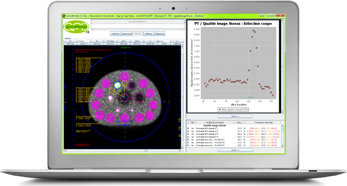
- Image quality on a thorax equivalent phantom2: Contrasts obtained on hot and cold fillable spheres relative to the medium in which they are situated (background)– Over-contrast coefficients for the hot spheres, in other words the ratios of contrast measured in the image relative to the intrinsic contrast of the object given by the ratio of the radioactive concentration between the spheres and the background – Residual error in scatter and attenuation corrections in the lung insert – Noise background Signal Ratio and Background signal variability associated with hot spheres, cold spheres and the lung insert.
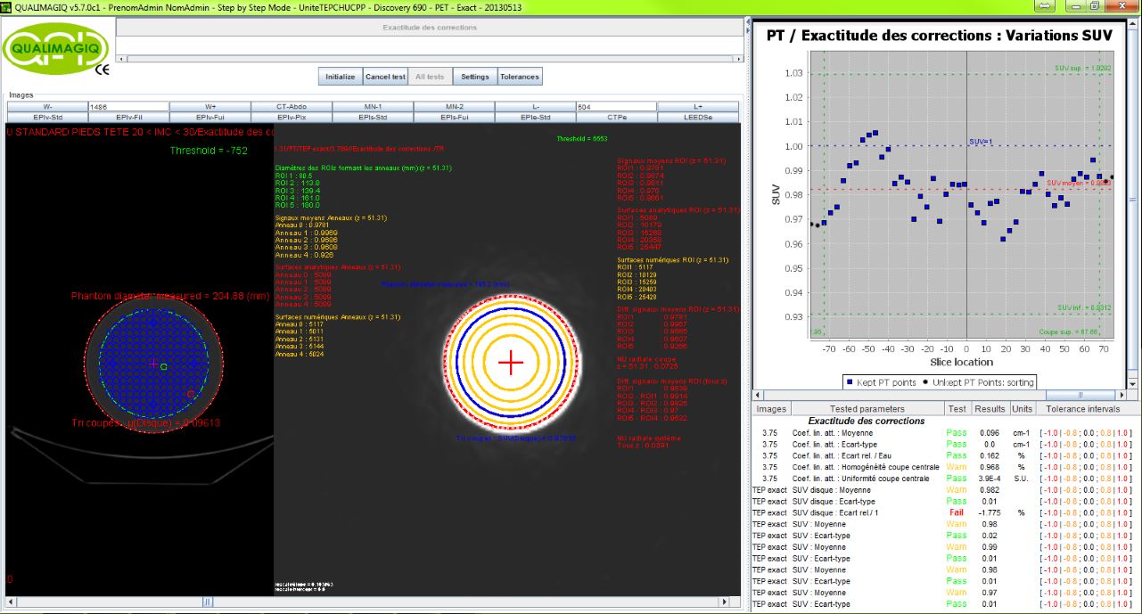
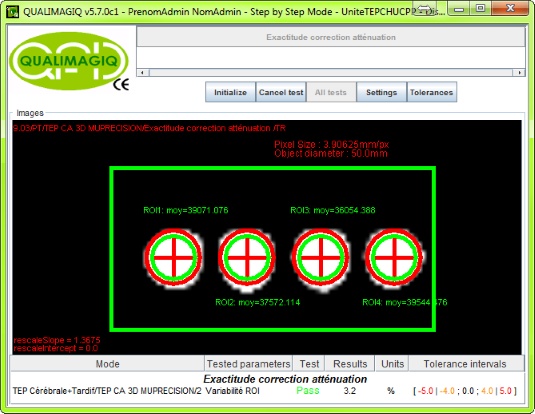
- Accuracy of corrections3: Control in the case of a homogeneous distribution of the radioactive concentration, of the quality of all of the corrections and of all of the calculations performed on the signal measured by the PET camera to obtain tomographic images calibrated in SUV. Thus on the CT images of a connected scanner, measurement of the average linear attenuation coefficient of water at 511 KeV, comparison to its theoretical value, study of its dispersion between slices and inside of the central slice of the cylindrical phantom. On the PET images, measurement of average SUV, comparison to its theoretical value of 1, study of its dispersion between slices and its radial distribution inside of these slices.
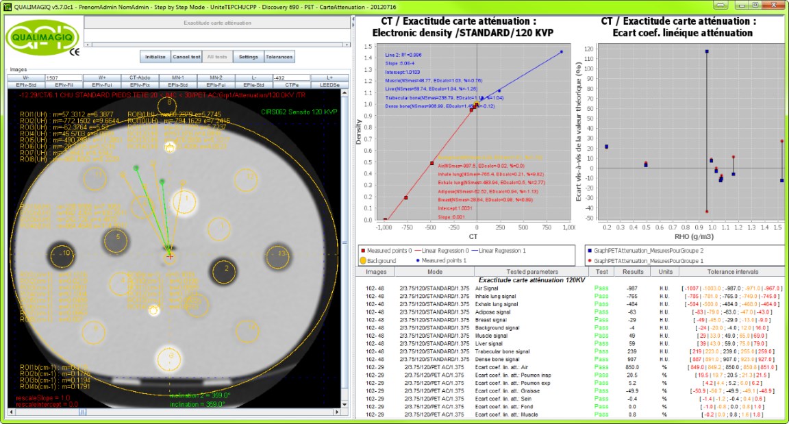
- Accuracy of the attenuation maps4: Indirect control of the quality of the corrections of attenuation originating from the scanner connected to the PET camera by measuring in the scanner images recomputed at 511 KeV by the scanner console (attenuation map), the linear attenuation coefficients (µ511)of a large range of reference materials whose coefficients are known to be 511 KeV. QUALIMAGIQ also enables you to complete this test by analysing in a similar manner the original scanner images obtained at 120 KV in order to distinguish a deviation between the measured µ and the reference µ already present in the original scanner at 120 KV from a deviation present solely in the scanner recomputed at 511 KeV. Incidentally, QUALIMAGIQ supplies based on the images of this original scanner a tracking of the stability of the conversion (Electron Density # Hounsfield units) and of the efficient energy of the X-Ray beam used to acquire the images.
- Accuracy of attenuation corrections5: This test is complementary to the Accuracy of the attenuation maps test on the attenuation maps produced by the scanner linked to a PET, however here QUALIMAGIQ tests whether these attenuation maps efficiently correct the attenuation of annihilation photons by verifying that the signals calibrated in raw radioactive or normalised (SUV) concentration are the same in a series of 2 to 6 test tubes containing the same radioactive concentration but different electron densities.
- Geometric alignment of the CT scanner on the PET imager6: Magnification deviation – Angular magnification deviations – Deviation in the position in the 3 directions of space – of a rectilinear source acquired in the 2 modalities.
Specifications
This module optimally fulfils all recommendations (rapidly, accurately and completely) put forth by the North American Association "National Electrical Manufacturer Association" (NEMA Report NU 2-2007) and by the French Medical Physics Association (SFPM Report N°24 2008). It automatically produces 6 different PDF analysis reports and a trend curve for each parameter tested.
Associated test objects:
- (1): 2 or 3 quasi-point radiation sources;
- (2): "Body" phantom specified in the 2007 NEMA-NU-2 publication (figures 7.1 page 31, 7.2 page 32 and 7.4 page 33) but also in the 2013 IEC 61675-1 publication, or "Thorax" phantom Elliptical Lung-Spine Body Phantom™;
- (3): hollow fillable cylinder Æ 10 cm whose length is greater than the axial FOV;
- (4): CIRS 062 or 062M phantom;
- (5): 2 to 5 cylindrical test tubes with waterproof seals;
- (6): line radiation source whose length is at least equal to half of the PET camera’s axial field of view.
Related products
Resources
You need a user manual? please contact us.
FAQ
Your brochure request has been added to your Download Cart.
At the end of your visit:
- Please click the "YOUR DOWNLOADS" button at the top right,
- Complete the form and SEND it.
You will get the documents shortly by email.