
Automates and Organises Quality Assurance in Radiation Therapy and Medical Imaging

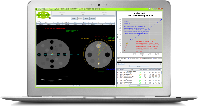
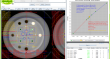
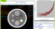
Connected to the QUALIMAGIQ platform, it takes the DE module only 2 mouse clicks to analyse all dicom images (tomographic slices) obtained with the CIRS 062 phantom, to construct the conversion curves scanner number – electronic density, and to control their constancy with regards to those registered in your TPS
Automatic electronic density calibration of images originating from a CT-scanner dedicated to radiation therapy or an “on-board” imager on a radiation therapy treatment machine with IGRT option used in tomographic mode (CBCT, kV or MV)
These automated analyses cover:
- Images coming from different voltages of the X- ray tube (kV) or from other MV energies;
- Images coming from 2 phantom diameters.
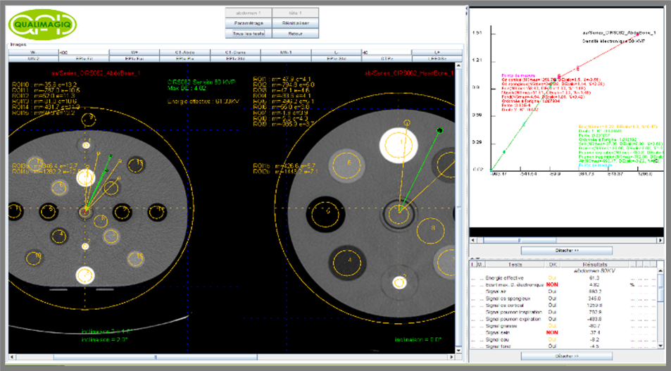
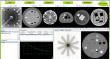
All of the measurement masks are realigned onto the images of the phantom (translations and rotations).
The dense signals (trabecular bone, cortical bone and titanium) can be extracted separately to avoid their “artifacting” onto weak and medium signals (air to soft tissue cavities). Of course the user may, if he or she wishes, also extract the bone and the soft tissues as well as the air cavities from one single and same image.
It is only mandatory for the titanium to be tested in a separate image because of the artefacts which it produces.
In addition to the 6 PDF reports, for the DE module QUALIMAGIQ creates a comprehensive report in MS-Excel file format, with a separate spreadsheet for each anatomy-energy pair.
This greatly simplifies entering of calibration data into your treatment planning system.
Specifications
Less than 10 minutes is all the time you need to perform the entire control: installation of the test object, image acquisition and analysis, editing of 6 different PDF analysis reports, 1 MS-Excel report and a trend curve for each tested parameter.
This module enables you to optimally fulfil requirements of the French National Agency for the Safety of Medicines (ANSM) decision dated 28/02/23 regulating the procedures for quality control of external radiation therapy and radiosurgery devices: point 8.1 of the annex.
Associated test objects:
- 062, 062M or 062MA from CIRS and GAMMEX 467 from SUN NUCLEAR.
Related products
FAQ
What are the necessary precautions to be taken to measure correctly the different CT numbers of human tissue equivalents if you also use a titanium insert?
Firstly, the titanium insert placed at the center of the phantom causes significant artifacts which disturb measurements performed in less dense inserts. It is therefore necessary to estimate the CT number of titanium insert on a image serie dedicated in which we do not measure the CT numbers of the other inserts less dense. These less dense insert will be measured in images of the phantom in which the titanium insert will replaced by an insert filled with water.
Secondly, it is important to work with the extended UH scale if you want to evaluate correctly the CT number corresponding to the titanium. The advantage of this scale is also to separate correctly in CT images the bony structures and hip prostheses. However images will take more space on hard disk because the signal will be coded on 16 bits against 12 bits.
Is it essential to align perfectly the density phantom in the CT-scanner before to make acquisition?
No for 2 reasons:
1- QUALIMAGIQ detects automatically phantom edges and its orientation before to extract the signal in the different inserts. Therefore QUALIMAGIQ compensates an eventual transversal (Up/Dowm and Left/right) misalignment.
2- The longitudinal alignment of the phantom is not essential because its tickness is 40 mm.
Your brochure request has been added to your Download Cart.
At the end of your visit:
- Please click the "YOUR DOWNLOADS" button at the top right,
- Complete the form and SEND it.
You will get the documents shortly by email.